POCUS – EMERGENCY ULTRASOUND
There are various names to describe these procedures. They involve scans of the abdomen, thorax and heart to give rapid information about a critical patient in a non-invasive low stress manor.
· POCUS – Point of Care Ultrasound Scan
- A general term that includes cardiac, thoracic and abdominal scans
· AFAST & TFAST – Abdominal & Thoracic Focused Assessment with Sonography for Trauma, Triage and Tracking
· Focused cardiac ultrasonography (FCU)
POCUS BASIC USE
- As a quick screen for potentially life-threatening problems:
1. Is there an abdominal effusion?
- The scan can identify the effusion, narrow down differentials, and locate appropriate windows for abdominocentesis
2. Is there free fluid in the pleural space?
- Provide oxygen and performs a thoracocentesis asap
- (Unless a coagulopathy related haemothorax is suspected in which case assess clotting first, e.g. rat bait cases)
3. Is there a pericardial effusion?
- If the patient is unstable this needs draining asap
4. Is there interstitial or alveolar fluid? (wet lung)
- If so, is the left atrium enlarged which could make cardiac causes more likely?
- If the assessment of the left atrium is not possible this judgement can be made via the clinical exam
o Heart murmur, arrythmia, pulmonary crackles etc. may increase the suspicion of left sided congestive heart failure as can the response to frusemide (radiographs to confirm when more stable)
o An abnormal Pro BNP blood test can also help the clinician suspect a cardiac cause of dyspnoea over a respiratory cause if ultrasound is unavailable
5. Is there a pneumothorax or consolidated lung?
COMMON INDICATIONS FOR A POCUS
· Collapsed/Critical patients:
- Screen for free fluid in the abdominal, pleural and pericardial space. Free fluid can be found in almost three quarters of unstable critical cases
- Facilitate guided abdo/thoracocentesis where appropriate for treatment & diagnostics
· Trauma patients:
- Free fluid screening – haemoabdomen, uroperitoneum. Track changes over time
- Pneumothorax (lack of glide sign) etc.
· Dyspnoea:
- Identification of pleural effusions
A very common cause of dyspnoeic cats
Can help to narrow down differentials
- Identify alveolar or interstitial fluid “wet lung”
Common ddx pulmonary oedema, pneumonia, neoplasia etc
- Confirm suspected L-CHF
Wet lung plus enlarged LA
- Identify consolidated lung – pneumonia, neoplasia
- Pneumothorax
Lack of glide sign
Altered M-mode
Note – Radiography is a superior modality of assessing the lungs but is often not safe to do initially in dyspnoeic patients. POCUS can give you useful information in the meantime to help guide diagnostics and treatment
Coagulopathy cases:
- Screen for free fluid - haemothorax/abdomen
Acute abdomen cases
Post-surgical unstable patients:
- Free fluid which could indicate bleeding or peritonitis etc.
Certain anaemic patients:
- Identify internal bleeding, obvious masses, organomegaly etc.
Urethral obstruction/urinary cases:
- Screen for uroperitoneum etc.
HOW TO DO A POCUS
Stabilise where appropriate first:
- Oxygen
- IV line and fluids
- Thoracocentesis - can be performed blind if there is a high suspicion of pneumothorax/pleural effusion from the exam
Patient prep:
May need to clip vs alcohol +/- gel and part the hair only.
Positioning:
Sternal/standing for any dyspnoeic patient.
Otherwise, lateral recumbency could be used.
T-FAST
1. Left and right chest tube sites
o Approx 7th to 9th intercostal spaces
o Place the probe on perpendicular to the chest wall
o For pulmonary parenchymal disease, oedema etc.
o Pneumothorax assessment
2. Left and right pericardial sites
o Approx 5th – 6th intercostal space
o Pericardial effusion assessment
o Pleural fluid assessment
o Basic assessment of left atrial size on the right
3. Diaphragmatic-hepatic window
o Place probe at the xiphoid and point cranially. Zoom out to image heart, pericardium and pleural space for fluid
NORMAL VS ABNORMAL PATHOLOGY
1. Left and right chest tube sites
“Normal view” – A-lines and glide sign:
A-LINES – Horizontal lines – pleural line reverberation artefact
· Seen with normal lung parenchyma if there is a normal “glide sign”
- The glide sign is the normal motion of the pleural line back and forth as the patient breathes
· If there are A-lines but no glide sign, the patient could have a pneumothorax
- Test thoracocentesis to confirm and treat
- There are M-mode assessments that can be performed to aid in pneumothorax identification but are beyond the scope of this introduction to POCUS section
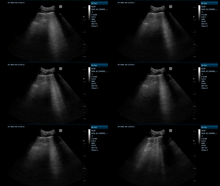
B-LINES – Vertical lines (“Rocket-lines”/” Wet-lung”)
- Vertical hyperechoic lines
- Indicate interstitial-alveolar pulmonary pathology. Ddx:
o Pulmonary oedema
o Non-cardiogenic pulmonary oedema – Includes haemorrhage, neoplasia, acute respiratory distress syndrome (ARDS) etc.
o Pneumonia
o Contusion etc.
- Occasional B-lines are normal
- 3 or more B- lines at one site is abnormal
Above cine loop - Normal A lines and glide sign
Above cine loop - Multiple B-lines
CONSOLIDATED LUNG
· Fluid or cells infiltrating the lung from pneumonia, pulmonary neoplasia, lung contusion or torsion
· Instead of normal A-lines - Hyperechoic foci with distal shadowing in early mild cases to “hepatised lung” in more advanced severe cases where the lung appears similar to the liver – Air filled bronchi show as hyperechoic air filled bronchograms and small hyperechoic foci represent air in the alveoli
· The lung retains its normal volume and shape unlike in atelectasis where the lung volume is reduced – often due to pressure from a PLE for example
Left and right pericardial sites
- Basic assessment of heart on the right - see focus cardiac ultrasound section
o Is the left atrium enlarged?
o Is the heart enlarged in general?
o Is the contractility subjectively reduced?
- Pericardial effusion assessment
- Pleural fluid assessment
PLEURAL EFFUSIONS
Pleural effusions can be picked up at the pericardial sites/caudoventral thorax
The above image shows a pleural effusion in a cat with heart failure (black anechoic fluid). Fibrin strands can be seen floating in this fluid.
Pleural effusion differentials:
· Cardiac – right sided congestive heart failure, pericardial effusion, cardiomyopathy secondary to hypertrophic cardiomyopathy or hyperthyroidism in cats
· Neoplasia
· Haemothorax – trauma, coagulopathy
· Pyothorax
· Pleuropneumonia
· Diaphragmatic hernia
· Hypoproteinaemia
· FIP
Oxygen should be provided and urgent thoracocentesis should be performed. Post drain radiographs screen for causes such as pulmonary masses. The POCUS (or a pro-BNP if ultrasound is unavailable) may help identify a cardiac cause. Fluid analysis and bloodwork including T4 (cats) should also be performed. See the dyspnoeic cat and pleural effusions section
PERICARDIAL EFFUSIONS
Screen for at the left and right pericardial sites (in the axillae) and also via the diaphragmatic hepatic view


